
New paper: Anterior eye surface changes following miniscleral contact lens wear
The prescription of scleral
contact lenses, as well as the number of practitioners who fit scleral contact
lenses, has notably increased over the last few years. The purpose of this work
was to quantify
the effect of short-term miniscleral contact lens wear on the anterior eye
surface of healthy eyes, including cornea, corneo-scleral junction and
sclero-conjuctival area. Twelve
healthy subjects wore a highly gas-permeable miniscleral contact lens of 16.5
mm diameter during a 5-hour period. Corneo-scleral height profilometry was
captured before, immediately following lens removal and 3 h after lens removal. Results
indicate that short-term miniscleral lens wear significantly modifies the
anterior eye surface. Significant planar limbal radius increment and flattening
in the sclero-conjuctival area were observed immediately following lens
removal. In addition, sclero-conjuctival flattening was not uniformly
distributed across the anterior eye, as the figure indicates.
Consejo A, Behaegel J, Van Hoey M, Wolffsohn JS, Rozema JJ, Iskander DR.
Anterior eye surface changes following miniscleral contact lens wear. 2018. Contact Lens and Anterior Eye (DOI:
doi.org/10.1016/j.clae.2018.06.005)
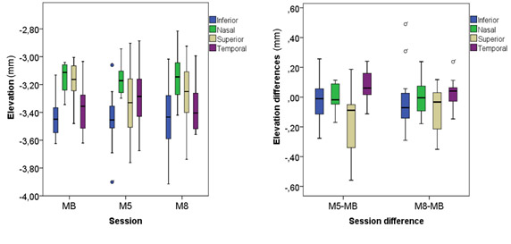
Figure. Left: Sclero-conjuctival elevation within
sectors for each session. Right: Difference respect to baseline in sclero-conjuctival
elevation within sectors. Sessions: Before contact lens wear (MB), immediately
after contact lens removal (M5) and 3 h after contact lens removal (M8). Error bars
indicate +/- one standard deviation; N=12.